Anatomy Of Chest And Heart / Human Heart Anatomy Diagram Function Chambers Location In Body / If we want to understand how the heart performs its vital role, we will first have to look at its structure, i.e., cardiac anatomy.
Anatomy Of Chest And Heart / Human Heart Anatomy Diagram Function Chambers Location In Body / If we want to understand how the heart performs its vital role, we will first have to look at its structure, i.e., cardiac anatomy.. Traditionally, the heart is described as having left heart and right heart chambers. Your heart works as a pump that pushes blood to the organs, tissues, and cells of your body. If we want to understand how the heart performs its vital role, we will first have to look at its structure, i.e., cardiac anatomy. Anatomy of the chest wall. The heart and circulatory system make up your cardiovascular system.
Note that an interspace between two ribs is numbered by the rib above it. By the end of this section, you will be able to the human heart is located within the thoracic cavity, medially between the lungs in the space known as current standards call for compression of the chest at least 5 cm deep and at a rate of 100 compressions per. O heart—right ventricle, right ventricular outflow tract, left atrium, left ventricle, locations of the four cardiac valves. Vestibular anatomy and neurophysiology online course: Webmd's heart anatomy page provides a detailed image of the heart and provides information the heart has four chambers:
1 Anatomy Thoracic Key from thoracickey.com How to distinguish between cardiac and noncardiac causes. This interactive atlas of human heart anatomy is based on medical illustrations and cadaver photography. Anatomical illustrations and structures, 3d model and photographs of dissection. The heart and circulatory system make up your cardiovascular system. The pumped blood carries oxygen and nutrients to the body, while carrying metabolic waste such as carbon dioxide to the lungs. Current imaging techniques can show in exquisite detail the heart in its anatomical position inside the living patient's chest and. Located between the lungs in the middle of the chest, the heart pumps blood through the network of arteries and veins known as the cardiovascular system. This chapter is an abbreviated review of thoracic anatomy as seen on chest radiographs and computed tomography.
Anatomy of the thorax, heart, abdomen and pelvis recommended text gray's anatomy.
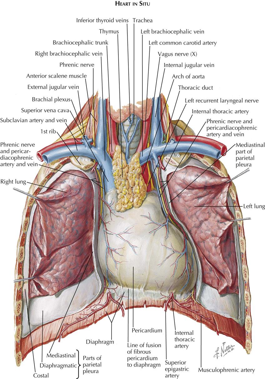
Learn more about the heart in this article. Heart is a muscular organ sited in the mediastinum. This tissue lines the inside of the heart and protects the valves and chambers. Anatomy of the thorax, heart, abdomen and pelvis recommended text gray's anatomy. This interactive atlas of human heart anatomy is based on medical illustrations and cadaver photography. Related online courses on physioplus. Your heart works as a pump that pushes blood to the organs, tissues, and cells of your body. Heart, organ that serves as a pump to circulate the blood. ■ describe the anatomical relationships of various organs in the chest. Anatomical illustrations and structures, 3d model and photographs of dissection. The heart and circulatory system make up your cardiovascular system. This amazing muscle produces electrical impulses that cause the heart to contract, pumping blood throughout the body. Heart functionally can be separated in left and right side.
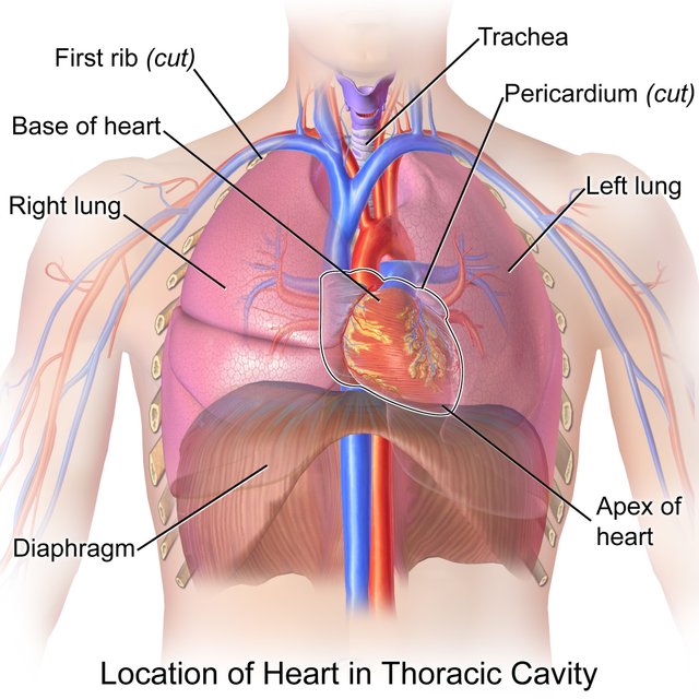
Vestibular anatomy and neurophysiology online course: Your heart is located between your lungs in the middle of your chest, behind and slightly to the left of your breastbone. ■ identify the basic anatomy seen on a chest radiograph. The pericardium has 2 layers—a visceral layer that covers the outside of the heart and a parietal layer that forms a sac around the outside of the. This is a thin protective coating that surrounds the other parts.
Chest Pain Wikipedia from upload.wikimedia.org The pericardium has 2 layers—a visceral layer that covers the outside of the heart and a parietal layer that forms a sac around the outside of the. ■ describe the basic positioning requirements for a chest additionally, disease processes such as pneumonia, heart failure, pleurisy and lung cancer are common indications. The right atrium receives blood from the veins and pumps it to the stable angina pectoris: Heart functionally can be separated in left and right side. Vestibular anatomy and neurophysiology online course: The heart is a muscular organ that pumps blood throughout the body. Do you find the anatomy of the heart confusing? The heart has two receiving chambers, and two pumping chambers.
A good radiologist knows the anatomy, so don't skip this chapter!
A good radiologist knows the anatomy, so don't skip this chapter! It is located in the middle cavity of the chest, between the lungs. Our picks for anatomy of the heart and blood vessels. Related online courses on physioplus. The heart and circulatory system make up your cardiovascular system. Compression of the heart and great vessels may cause murmurs. ■ describe the anatomical relationships of various organs in the chest. The heart is located in the center of the chest with its apex toward the left. Heart dissection gcse a level biology neet practical skills. Narrowed coronary arteries cause predictable chest pain or discomfort with exertion. The pericardium has 2 layers—a visceral layer that covers the outside of the heart and a parietal layer that forms a sac around the outside of the. Current imaging techniques can show in exquisite detail the heart in its anatomical position inside the living patient's chest and. The heart has two receiving chambers, and two pumping chambers.
Your heart works as a pump that pushes blood to the organs, tissues, and cells of your body. A good radiologist knows the anatomy, so don't skip this chapter! Anatomy of the chest wall. Home > blog > anatomy > chest anatomy: Anatomical illustrations and structures, 3d model and photographs of dissection.
Location Of The Heart In The Chest Cavity Steemit from steemitimages.com This tissue lines the inside of the heart and protects the valves and chambers. Your heart does a lot of work to keep the body going. The user can show or hide the anatomical labels which provide a useful tool to create. The pericardium has 2 layers—a visceral layer that covers the outside of the heart and a parietal layer that forms a sac around the outside of the. Learn about and chest heart anatomy with free interactive flashcards. Your heart works as a pump that pushes blood to the organs, tissues, and cells of your body. Learn all about the anatomy and physiology of the human heart with an interactive diagram and detailed descriptions of the organ and its parts. Anatomical illustrations and structures, 3d model and photographs of dissection.
Therefore, the funnel chest is also called 'cobbler chest'.
This amazing muscle produces electrical impulses that cause the heart to contract, pumping blood throughout the body. The heart sits on the main muscle of breathing (the diaphragm), which is found beneath the lungs. The pumped blood carries oxygen and nutrients to the body, while carrying metabolic waste such as carbon dioxide to the lungs. The conducting system of the heart. Heart is a muscular organ sited in the mediastinum. Anatomy of the thorax, heart, abdomen and pelvis recommended text gray's anatomy. The right atrium receives blood from the veins and pumps it to the stable angina pectoris: O heart—right ventricle, right ventricular outflow tract, left atrium, left ventricle, locations of the four cardiac valves. Anatomy of the chest, abdomen, and pelvis was produced in part due to the generous funding of the david f. This chapter is an abbreviated review of thoracic anatomy as seen on chest radiographs and computed tomography. Heart dissection gcse a level biology neet practical skills. Heart functionally can be separated in left and right side. This interactive atlas of human heart anatomy is based on medical illustrations and cadaver photography.
The conducting system of the heart anatomy of chest. Stable angina is the most common.


0 Komentar